For research use only. Not for therapeutic Use.
Pexidartinib (PLX-3397) is a potent, orally active, selective, and ATP-competitive colony stimulating factor 1 receptor (CSF1R or M-CSFR) and c-Kit inhibitor, with IC50s of 20 and 10 nM, respectively. Pexidartinib (PLX-3397) exhibits 10- to 100-fold selectivity for c-Kit and CSF1R over other related kinases. Pexidartinib (PLX-3397) induces cell apoptosis and has anti-tumor activity[1].
Pexidartinib (PLX-3397) is a potent, selective and ATP-competitive CSF1R (cFMS) and c-Kit inhibitor, shows 10- to 100-fold selectivity for c-Kit and CSF1R over other related kinases, such as FLT3, KDR (VEGFR2), LCK, FLT1 (VEGFR1) and NTRK3 (TRKC), with IC50s of 160, 350, 860, 880, and 890 nM, respectively[1].
Pexidartinib can be used to deplete the microglia cells in mice.
Pexidartinib (290 ppm in AIN-76A standard chow, 21 days) depletes the microglia cells in brain by 70% in adult male C57BL/6 J mice (20–25 g)[5].
Pexidartinib (600 ppm in chow, 10 days and 30 days ) depletes the microglia cells more than 90% in brain of C57BL/6J mice[6].
Pexidartinib (PLX3397; 0.25, 1 mg/kg, twice daily for 8 days) inhibits the proliferation of microglia and BrdU-positive cells in neonatal mice[2].
Pexidartinib (1 mg/kg, twice daily for 8 day) shows no obvious effect on the cleaved caspase-3-positive cells in mice[2].
Pexidartinib (50 mg/kg; p.o.; every second day for 3 weeks) reduces tissue macrophage levels without affecting glucose homeostasis in mice[4].
| CAS Number | 1029044-16-3 |
| Synonyms | 5-[(5-chloro-1H-pyrrolo[2,3-b]pyridin-3-yl)methyl]-N-[[6-(trifluoromethyl)pyridin-3-yl]methyl]pyridin-2-amine |
| Molecular Formula | C20H15ClF3N5 |
| Purity | ≥95% |
| InChI | InChI=1S/C20H15ClF3N5/c21-15-6-16-14(10-28-19(16)29-11-15)5-12-2-4-18(26-7-12)27-9-13-1-3-17(25-8-13)20(22,23)24/h1-4,6-8,10-11H,5,9H2,(H,26,27)(H,28,29) |
| InChIKey | JGWRKYUXBBNENE-UHFFFAOYSA-N |
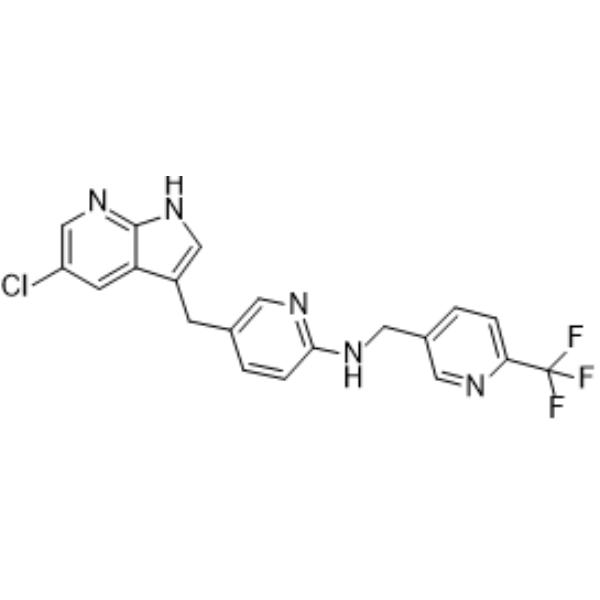
| SMILES | C1=CC(=NC=C1CC2=CNC3=C2C=C(C=N3)Cl)NCC4=CN=C(C=C4)C(F)(F)F |
| Reference | [1]. DeNardo DG, et al. Leukocyte complexity predicts breast cancer survival and functionally regulates response to chemotherapy. Cancer Discov. 2011 Jun;1(1):54-67. [2]. Kuse Y, et al. Microglia increases the proliferation of retinal precursor cells during postnatal development. Mol Vis. 2018 Jul 30;24:536-545. eCollection 2018. [3]. Lee JH, et al. A phase I study of pexidartinib, a colony-stimulating factor 1 receptor inhibitor, in Asian patients with advanced solid tumors. Invest New Drugs. 2019 Mar 2. [4]. Merry TL, et al. The CSF1 receptor inhibitor pexidartinib (PLX3397) reduces tissue macrophage levels without affecting glucose homeostasis in mice. Int J Obes (Lond). 2020;44(1):245-253. [5]. Luo L, et al. Intermittent theta-burst stimulation improves motor function by inhibiting neuronal pyroptosis and regulating microglial polarization via TLR4/NFκB/NLRP3 signaling pathway in cerebral ischemic mice. J Neuroinflammation. 2022 Jun 11;19(1):141. [6]. Chadarevian JP, et al. Engineering an inhibitor-resistant human CSF1R variant for microglia replacement. J Exp Med. 2023 Mar 6;220(3):e20220857. |
| Chemistry Calculators | Dilution Calculator In vivo Formulation Calculator Molarity Calculator Molecular Weight Calculator Reconstitution Calculator |